
An ingrown toenail is a condition that occurs when part of a nail injures the surrounding skin. This problem particularly affects the big toe.

An ingrown toenail is a condition that occurs when part of a nail injures the surrounding skin. This problem particularly affects the big toe.

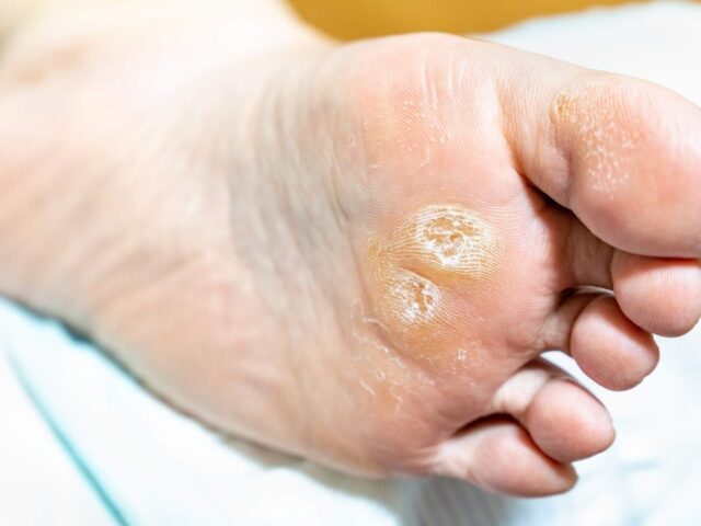
Corns on the foot: relieve this localized foot pain
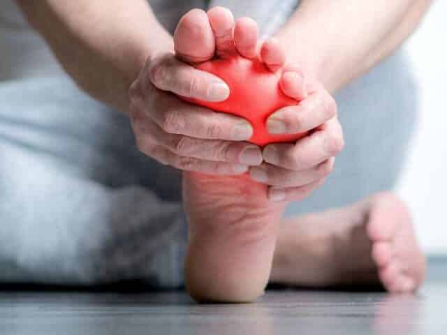
You feel a sharp pain on the top of your second toe. Every step in your shoes triggers that nagging discomfort. You discover a small circular area of hard, yellowish skin. This painful prominence forces you to change your gait.
Or maybe that feeling of having a stone in your shoe between your toes persists despite repeated checks. This localized pain interferes with your daily activities.
You probably suffer from corns, these localized accumulations of thickened skin that create painful pressure on the underlying tissues.

A corn is a localized and concentrated thickening of the surface layer of the skin. Unlike calluses that extend over wider areas, corns grow on a specific point of friction or repeated pressure.
This keratin buildup forms a conical central nucleus that penetrates inward, compressing the underlying tissues. This inverted cone-shaped structure explains the characteristic pain — like stepping on a nail or a thorn.
Hard corns (heloma durum) appear on surfaces exposed to dry friction. The tops of the toes, the prominent joints, the outer sides of the fifth toes — these areas are subject to the constant friction of the shoes. The skin becomes thick, hard and yellowish.
Soft corns (heloma molle), commonly known as partridge’s eye, develop between the toes in a moist environment. Maceration makes the horn whitish and spongy rather than hard. This variety nevertheless causes severe pain, amplified by the sensitivity of the interdigital skin.
Subungual corns form under the toenails, creating painful pressure between the nail and the nail bed. This particular location complicates self-processing.
Toe deformities are the main cause. Hammer, claw, or mallet toes create bony prominences that rub against the shoes. These angulated joints are under constant pressure, stimulating the formation of corns.
Unsuitable shoes make the problem much worse. Shoes that are too tight that compress the toes. Rough inner seams that irritate the skin. Shoes with a pointed toe that force the toes into unnatural positions. These mechanical factors keep the friction responsible for corns.
Hallux valgus (bunion) pushes adjacent toes against each other, creating interdigital pressure points. This deformity particularly favours interdigital corns.
Biomechanical abnormalities change the distribution of pressures during walking. A foot that pronounces excessively or has an arch that is too high alters the normal mechanics, creating areas of abnormal friction.
Professional activity also influences the development of corns. Occupations that require repeated kneeling positions or the wearing of rigid safety shoes increase the risks.
This confusion occurs frequently. Corns and plantar warts both appear as areas of painful skin thickening. However, their nature differs fundamentally.
A corn is the result of mechanical pressure. The plantar wart comes from a viral infection (human papillomavirus). On close examination, the wart has small black dots (thrombosed blood vessels). The horn shows a translucent central core without these characteristic points.
The pain also differs. The horn hurts when the vertical direct pressure is used. The wart causes pain during lateral compression (pinching). This distinction helps to guide the diagnosis.

At Médecine podiatrique du Plateau, Drs. Sandra Gendron, Dr. Stephen Davis and Dr. Émile Carrier, podiatrists, treat corns with a comprehensive approach.
Podiatric debridement meticulously removes the corn. We use sterilized scalpel blades to enucleate the central nucleus. This precise extraction immediately relieves painful pressure. The procedure, which could seem impressive, is painless when performed correctly on thickened skin.
For a corn between the toes, we reduce the skin thickness and sometimes place toe separators. These foam or silicone devices maintain proper spacing, reducing the friction that causes recurrence.
Subungual corns often require thinning or reduction of the nail to remove pressure on the underlying corn.
Beyond symptomatic relief, we identify structural causes. Biomechanical examination reveals pressure imbalances. The analysis of your toe deformities guides therapeutic recommendations.
Custom-made foot orthotics correct biomechanical abnormalities contributing to corns. By optimising pressure distribution, these devices reduce the mechanical stresses responsible.
Specialized protective pads cushion vulnerable areas. Gel tubes to protect toes. Foam dressings to reduce friction. Felt rings surrounding the horn to redistribute pressure. These accessories extend the relief between professional treatments.
Recommendations for the right footwear are crucial. Sufficient toe box height to accommodate deformations. Suitable width to avoid compression. Soft materials that do not create friction points.
Dr. Émile Carrier, podiatrist and co-owner of our clinic, emphasizes: “Over-the-counter products containing salicylic acid carry risks, especially for people living with diabetes or with compromised circulation. These products dissolve the skin indiscriminately, creating chemical burns on adjacent healthy tissue. The professional intervention precisely removes the corn without damaging the surrounding skin. »

Corns will inevitably recur if the mechanical causes persist. Debridement offers temporary relief, but without correction of the causal factors, the corn reforms within a few weeks.
Wear appropriate footwear that offers generous space for your toes. Avoid shoes with pointed toes and high heels that compress the forefoot.
If you have toe deformities, consult a doctor to evaluate corrective options. Orthotics, toe separators, and in some cases, corrective surgery may be necessary for lasting resolution.
If your corns cause pain that limits your activities, if you observe signs of infection (redness, heat, drainage), if you live with diabetes (never try self-treatment), or if corns recur quickly despite your precautions, consult for a professional evaluation.
For the treatment of your corns and calluses, contact us at 819-800-1212 or fill out the contact form to schedule an appointment.
Podiatric consultations are NOT covered by the RAMQ. Private insurance can cover our services. Find out about your coverage before you show up. We do not do direct billing.
Calluses on the feet: professional care to regain comfort
You put on your shoes in the morning. That unpleasant feeling under the forefoot reminds you of its presence. Thickened and hardened skin makes it uncomfortable with every step. You try to file this horn with a pumice stone. The thickness decreases temporarily, but returns quickly, sometimes thicker than before.
These calluses that seem harmless often hide a biomechanical imbalance that requires professional intervention.

Calluses (hyperkeratosis) and calluses represent a thickening of the surface layer of the skin in response to repeated pressure or friction. Your body produces this protective fabric to defend your skin against excessive mechanical stress.
Calluses usually form on larger areas — the sole of the foot, heels, the base of the toes. This buildup of hardened skin develops gradually over weeks or months of repeated use.
Corns are a more localized and concentrated form of hyperkeratosis. These punctiform thickenings possess a hard central nucleus that penetrates the underlying tissues, creating acute pain on pressure. Corns frequently develop on the top of the toes or between the toes in reaction to the friction of shoes or toes against each other.
This skin response, although initially protective, paradoxically becomes problematic. The excessive thickness itself creates additional pressure. A vicious circle sets in: pressure causes thickening, which increases pressure, which stimulates more thickening.
Biomechanical imbalances are the main cause. A foot that collapses excessively (pronation,
Structural deformations concentrate pressures on specific points. Hallux valgus (bunion) transfers loads to the second and third metatarsal heads. Hammer toes create friction points on prominent joints. These architectural anomalies inevitably generate areas of hyperkeratosis.
Unsuitable footwear makes the problem worse, such as shoes that are too tight that compress, high heels that transfer excessive pressure to the forefoot, and worn shoes that have deteriorated cushioning. These factors amplify the mechanical stress on your skin.
Intense physical activity repeatedly solicits the same areas. Runners frequently develop calluses under the metatarsal heads. The dancers have characteristic thickenings depending on their discipline.
Aging can thin the plantar fat pad. This loss of natural protection exposes the skin to more pressure, promoting hyperkeratosis.
In healthy people, calluses are mainly a discomfort and aesthetic concern. However, excessive thickness can become painful and limit activities.
For people living with diabetes, calluses are a real danger. Under this thickened layer of skin, a hematoma can form without you feeling it if neuropathy has decreased your sensitivity. This pooled blood creates an environment prone to ulceration. A wound develops under the callus, invisible to the naked eye. This silent progression sometimes leads to serious infections even before the problem is detected. To learn more, read our article on diabetes-related foot problems and the Société des sciences vasculaires du Québec’s article on diabetic foot.
Cracks in calluses, especially in the heels, create entry points for infections. These deep, painful cracks bleed easily and heal with difficulty.

At Médecine podiatrique du Plateau, podiatrists Dr. Sandra Gendron, Dr. Stephen Davis and Dr. Émile Carrier offer comprehensive professional care to treat calluses and calluses.
Podiatric debridement meticulously removes layers of thickened skin. We use specialized sterilized instruments — scalpel blades, rotary burs — to gradually reduce hyperkeratosis. This professional technique is radically different from attempts at self-treatment with graters or pumice stones.
We work precisely to remove excess thickness without damaging the underlying healthy skin. This expertise protects your foot while maximizing relief. The result is immediate: you leave with visibly smoother and more comfortable feet.
For people living with diabetes, we thoroughly inspect under each callus. This vigilance detects invisible preulcerations on the surface. This early detection prevents the progression to complete wounds that would require complex interventions.
The treatment of heel cracks combines the debridement of surrounding calluses with the application of specialized emollient products. We prescribe creams containing molecules like urea in high concentrations (20-40%) that deeply moisturize and promote healing.
Beyond immediate relief, we identify the underlying causes of your calluses. Analysis of your biomechanics reveals pressure imbalances. The examination of your shoes detects aggravating factors.
Custom-made foot orthotics redistribute plantar pressure. These devices compensate for the biomechanical imbalances that cause your calluses. By normalizing load distribution, orthotics can reduce the rate of reformation of hyperkeratosis and relieve the associated discomfort as it develops.
We guide you in choosing the right shoes: the right width, enough cushioning and the right support. These features reduce the mechanical stress responsible for calluses.
Dr. Sandra Gendron, podiatrist, explains: “Professional debridement offers immediate relief, but without biomechanical correction, calluses return quickly. Our approach combines symptomatic treatment and correction of causes. This all-encompassing strategy keeps your feet comfortable in the long run. »
The periodicity of care varies according to the speed of reformation of your calluses. Some people receive care every six to eight weeks. Others, particularly those with significant deformities or intense physical activity, require monthly interventions.
People living with diabetes should receive regular professional podiatric care, even if calluses seem minimal. This preventive monitoring detects problems before they become serious.
If your calluses are causing discomfort, if you are experiencing painful cracks, if you are living with diabetes (routine consultation recommended), or if your attempts at self-treatment are ineffective or aggravating the problem, consult for professional care.
For professional care of your calluses and calluses, contact us at 819-800-1212.
Podiatric consultations are NOT covered by the RAMQ. Private insurance usually covers our services. Documentation systematically provided for your complaints.

Foreign body in the foot: safe extraction and prevention of complications
You walk barefoot at home. Sharp pain stops you in your tracks. You examine your foot. A small wound bleeds slightly. You try to see if what hurt you has lodged in your foot. You can’t find anything, but the walk is painful.
The days go by. The wound refuses to heal. Redness spreads around the site. The area becomes tender and swollen. This persistent pain prevents you from walking normally.
A foreign body lodged in your foot often requires professional extraction to avoid infectious complications and ensure complete healing.

Wood splinters are the most common type. These fragments easily penetrate the skin, especially at the sole of the foot where the skin thickness is important. Wood fragments easily during extraction attempts, leaving pieces deeply buried.
Glass shards occur when walking on debris. The glass is radiopaque, which makes it easier to locate by X-ray. Its smooth, sharp surface can penetrate deep into tissues without breaking.
Plant thorns (rose, cactus, hawthorn) often have prickles or hairs that anchor them in the tissues. This structure makes extraction difficult and promotes the inflammatory response.
Metal fragments (nails, staples, splinters) penetrate deep during piercing trauma. Rusty metal increases the risk of infection, including tetanus.
Localized pain that persists beyond a few days suggests the presence of a residual foreign body. This pain intensifies when pressing or walking.
Chronic inflammation manifests as persistent redness, swelling, and local warmth. The wound refuses to heal completely despite proper care.
Intermittent drainage of fluid or pus often indicates an infected foreign body. The organism attempts to expel foreign material through this process of drainage.
The formation of a cyst or subcutaneous mass is sometimes the result of the encapsulation of the foreign body by the tissues. This defensive reaction isolates foreign material but creates a palpable and sometimes painful mass.
Infection is the most common complication. The bacteria enter with the foreign body or secondarily colonize the wound. Without treatment, the infection can spread to deep tissues, tendons, or even bone.
Organic foreign bodies (wood, plant thorns) trigger intense inflammatory reactions. These materials break down in tissues, amplifying the immune response and prolonging inflammation.
Tetanus threatens when objects contaminated by soil penetrate deeply. This life-threatening infection warrants checking your vaccination status and updating it if necessary.
Damage to deep structures occurs when the foreign body reaches tendons, nerves, or blood vessels. These complications sometimes require specialized surgical management.

At Médecine podiatrique du Plateau, podiatrists Dr. Sandra Gendron, Dr. Stephen Davis and Dr. Émile Carrier safely extract foreign bodies under sterile conditions.
If necessary, we use local anesthesia for your comfort. An injection of lidocaine completely numbs the area, allowing for painless careful extraction.
If necessary, on-site X-rays locate radiopaque foreign bodies (glass, metal). This imaging guides the incision and extraction, minimizing tissue damage. Non-radiopaque materials (wood, plastic, plant thorns) require precise clinical localization and sometimes careful surgical exploration.
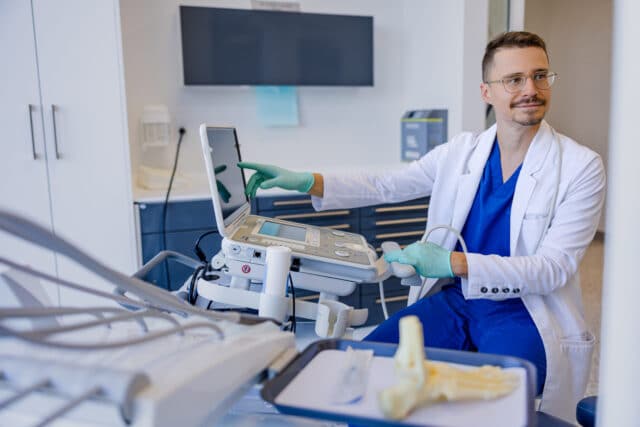
Musculoskeletal ultrasound visualizes certain non-radiopaque foreign bodies. This technology detects splinters and assesses the inflammatory response of the surrounding tissues.
After complete extraction, we clean the wound thoroughly. This pressure washing removes debris and reduces bacterial load. Irrigation is a crucial step in preventing infection.
If the infection has developed, we prescribe appropriate antibiotics. Deeply infected wounds sometimes require debridement (removal of necrotic tissue) to promote healing.
We check and update your tetanus protection as recommended. This precaution protects against a potentially serious complication.
Dr. Émile Carrier, podiatrist and co-owner of our clinic, warns against home treatments: “Attempts at home extraction often fragment the foreign body, complicating subsequent extraction. Early professional intervention avoids these complications and ensures complete removal under optimal conditions.”
Wear appropriate footwear in risky environments. Construction sites, yards with plant debris, rocky beaches: these situations warrant adequate protection.
Inspect your surroundings before walking barefoot. Check decks, lawns and floors for potential hazards.
Keep your outdoor spaces clean. Pick up wood debris, broken glass, and stray nails.
For people living with diabetes or neuropathy, barefoot walking is not recommended, even indoors. The loss of sensitivity delays the detection of foreign objects, allowing deep penetration before you notice the problem.
Consult a doctor quickly if you are unable to completely remove a foreign body, if the pain persists for more than a few days, if signs of infection appear (increasing redness, heat, purulent drainage), or if you are not sure if your tetanus vaccination is up to date.
For a safe removal of a foreign body from your foot, contact us at 819-800-1212 or fill out the contact form by clicking here.
Podiatric consultations are NOT covered by RAMQ. Private insurance usually covers our services.

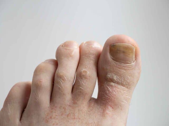
Athlete’s foot: treating and preventing this common fungal infection
You take off your socks at the end of the day. That persistent itch between your toes has been tormenting you for weeks. The skin whitens and peels. An unpleasant smell emanates from your feet despite rigorous hygiene.
You apply moisturizers. The irritation worsens. That redness that extends between your toes resists all your self-treatment efforts.
You probably suffer from athlete’s foot, the most common fungal infection affecting the feet. This skin fungus, medically called tinea pedis, affects millions of people every year.
Athlete’s foot results from infection with microscopic fungi (dermatophytes) that proliferate in warm, humid environments. These organisms feed on the keratin present in the superficial layer of your skin.
The name “athlete’s foot”, or tinea pedis, comes from the high frequency of this infection in athletes. Athletic shoes create an ideal environment for fungal growth. Heat, humidity and maceration: these conditions favour the rapid multiplication of fungi.
Contrary to popular belief, you don’t have to be an athlete to develop this infection. Anyone exposing their feet to prolonged moisture is at risk.
The infection typically affects the spaces between the toes, especially between the fourth and fifth toes. The interdigital form represents the most common type. The moccasin shape covers the sole and sides of the foot with a dry, flaky layer. The rarer vesicular form causes fluid-filled blisters.

Itching is the dominant symptom. This unpleasant feeling often intensifies after you take off your shoes or when your feet become wet.
The skin between your toes turns white and becomes soft (maceration). It flakes into small shreds. Painful cracks may appear in the interdigital spaces. These crevices create entry points for secondary bacterial infections.
Redness and inflammation border the affected areas. Dry, flaky skin sometimes extends over the sole and sides of the foot. The unpleasant odour results from the breakdown of skin tissue by fungi and the associated bacterial overgrowth.
The infection spreads easily to toenails, causing thickening, yellowish discoloration, and crumbling. This extension significantly complicates the treatment. This is nail fungus, or onychomycosis.

At Médecine podiatrique du Plateau, podiatrists Dr. Sandra Gendron, Dr. Stephen Davis and Dr. Émile Carrier diagnose athlete’s foot through a clinical examination. The characteristic appearance and typical location quickly guide the diagnosis.
Consulting one of our podiatrists is helpful in distinguishing this problem from other skin diseases such as eczema and psoriasis.
In atypical or treatment-resistant cases, we can take a skin sample for mycological confirmation in the laboratory. This analysis precisely identifies the responsible organism and guides the optimal therapeutic choice.
Treatment is mainly based on topical antifungals. Creams, powders, or sprays containing antifungals like ciclopirox olamine or terbinafine. Daily application for two to four weeks. It is important to treat for this duration to prevent recurrence rather than treating only until symptoms stop.
Your podiatrist can also prescribe specialized dressings that wick away moisture, get to the root of the problem while avoiding the disadvantage of using creams, which is to maintain a moist environment, especially between the toes.
For widespread, severe or recalcitrant infections, we can prescribe oral antifungals. These systemic medications reach the fungi that topical treatments do not penetrate properly. A treatment of several weeks may be necessary for complete eradication, especially if the nails are also affected.
If the nails are infected, their treatment becomes essential. Fungal nails serve as a reservoir of fungi that constantly reinfect the treated skin. We offer treatment options for nail fungus, including specialized topical treatments and oral antifungals.
Secondary bacterial infections sometimes require antibiotics. The deep cracks between the toes allow bacteria to seep in. This superinfection manifests itself in increased redness, purulent drainage and intensified pain.
Dr. Émile Carrier, podiatrist and co-owner of our clinic, explains that athlete’s foot frequently recurs if the factors that promote fungal growth persist. Treatment of active infection should be accompanied by rigorous preventive measures to avoid re-infection.
Keep your feet dry. Dry thoroughly between the toes after each wash or exposure to water. This step, which is often neglected, is your best protection.
Change your socks daily, more frequently if your feet are sweating profusely. Choose socks made of moisture-wicking fibers rather than cotton that retains it.
Alternate your shoes. Wear different pairs each day to allow for thorough drying. Wet shoes promote fungal growth.
Wear sandals in locker rooms, public showers and swimming pools. The moist surfaces of these environments are home to fungi that easily infect bare feet.
Apply preventive antifungal powders if you have a susceptibility to recurrent infections. This proactive approach significantly reduces recidivism.
Treat hyperhidrosis (excessive sweating) if you have it. Excessive moisture control protects against fungal infections. Your podiatrist may prescribe products to treat this problem.
If symptoms persist despite two weeks of over-the-counter treatment, consult one of our podiatrists. Resistant infections require stronger antifungals or investigation to rule out other skin pathologies.
The presence of signs of secondary bacterial infection — intense redness, heat, purulent drainage, fever — warrants a prompt assessment. These complications require antibiotic treatment.
People living with diabetes or immunosuppression should seek medical attention as soon as symptoms appear. In these vulnerable populations, a minor fungal infection can progress to serious complications.
If you have any questions about athlete’s foot or to make an appointment, contact us at 819 800-1212 or fill out the contact form.
Podiatric consultations are NOT covered by the RAMQ. However, several private insurance plans cover our services.

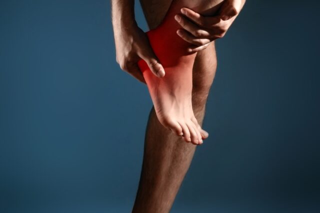
Ankle sprain: quick diagnosis and complete treatment in Gatineau
You walk on an uneven sidewalk. Your ankle twists sharply inwards. The shooting pain stops you immediately. You try to limp on your way. Swelling appears quickly.
You ask yourself, “Is this a big deal? Should I consult? Can I continue walking? »
Ankle sprains are one of the most common musculoskeletal injuries. Yet, many people minimize this injury and neglect proper treatment. This neglect often turns an acute sprain into chronic instability that persists for years.
At Médecine podiatrique du Plateau, Dr. Sandra Gendron, Dr. Stephen Davis and Dr. Émile Carrier, podiatrists evaluate and treat ankle sprains with advanced diagnostic technologies. Our clinic is located at the AGORA complex in Gatineau (Plateau sector). X-ray and musculoskeletal ultrasound on site. We offer you personalized care and treatment plan. The goal? A complete rehabilitation.
This guide helps you understand ankle sprain, recognize its severity, and discover treatment options that speed up your recovery.

A sprain occurs when the ligaments stabilizing your ankle are overstretched or torn. Ligaments are bands of strong connective tissue that hold bones together. Unlike muscles that actively contract, ligaments provide passive but essential support.
Lateral (external) ankle sprain accounts for 85% of cases. Your foot turns inward abruptly (inversion), stretching or tearing the ligaments on the outside of the ankle. The anterior talofibular ligament suffers the most frequent damage, followed by the calcaneofibular ligament.
Sprains are classified according to their severity.
Grade 1 (mild): ligament stretch without significant tearing, moderate pain, minimal swelling, ability to walk retained.
Grade 2 (moderate): partial tear of ligaments, marked pain, significant swelling, difficulty walking, mild instability.
Grade 3 (severe): complete tear of one or more ligaments, severe pain, massive swelling, inability to support weight, significant instability.
These terms refer to the same injury and are often used interchangeably. Sprains (or strains) affect the ligaments that connect the bones together. When your ankle twists, the ligaments overstretch or tear.
This terminological confusion sometimes stems from popular usage where some people use “strain” to describe a mild sprain and “sprain” for a more severe injury. In reality, it is the same type of ligament injury, only the severity of which varies.
Characteristic symptoms include pain, swelling, and functional difficulty. Clinical examination and imaging confirm the exact nature of your injury and determine its severity.
This question comes up constantly. The answer depends on the severity of your sprain.
A mild sprain (grade 1) usually allows you to walk with moderate discomfort. You’re probably limping but you can support your weight. This functional capacity suggests that the ligaments, although stretched, maintain sufficient stability.
A moderate sprain (grade 2) makes walking difficult and painful. You could technically walk but at the cost of considerable effort and pronounced lameness. This early mobilization may aggravate partial ligament tears.
A severe sprain (grade 3) makes walking without support usually impossible. The intense pain and marked instability prevent normal loading. Forcing the walk in this situation prolongs healing and risks further damage to already compromised structures.
Dr. Émile Carrier, podiatrist and co-owner of our clinic, explains: “The ability to walk immediately after the injury does not guarantee the absence of a significant ligament fracture or tear. On the other hand, if you are unable to put weight on the affected ankle immediately after the accident, this is a great reason to see for x-rays to check for a fracture. If a fracture is identified, the treatment will be very different from that of a sprain. »
The timeline of healing varies greatly depending on the severity of the sprain.
A mild sprain (grade 1) usually resolves in 1 to 3 weeks. The pain decreases quickly. The swelling gradually subsides. You are gradually returning to your normal activities.
A moderate sprain (grade 2) requires 3 to 6 weeks of recovery. The first few weeks often require protection with a splint or walking boot. Rehabilitation with amplitude and strengthening exercises becomes crucial to restore optimal function.
A severe sprain (grade 3) takes 8 to 12 weeks or more to heal completely. Strict initial immobilization protects torn ligaments while they heal. Gradual and supervised rehabilitation extends over several months to recover strength, proprioception and stability.
These delays represent the healing of the ligament tissues. Full functional recovery — including the return to demanding sports and activities — may require additional time.
Our on-site diagnostic capability speeds up your care and eliminates frustrating wait times.
Immediate digital radiography excludes associated fractures. Approximately 22% of ankle sprains are associated with fractures (references 1 and 2)—malleoli, base of the fifth metatarsal, posterior talus process. These fractures require separate management. You get this confirmation at your initial consultation.

Musculoskeletal ultrasound directly visualizes the injured ligaments. This technology evaluates the structural integrity of each ligament. It detects partial or complete tears. It identifies hematomas and periligament edema. This precise information guides our treatment recommendations.
The thorough clinical examination evaluates functional stability. Ligament stress tests. Joint range of motion assessment. Palpation of anatomical structures. This evaluation complements the imaging to establish the precise grade of your sprain.
Optimal healing is based on appropriate care from the first days.
The PEACE and LOVE protocol developed by physiotherapist Blaise Dubois and physiotherapist and researcher Jean-François Esculier has become our reference in the management of ankle sprains. The protocol is all about:
P – Protection : Protect the injured area by limiting movements that increase pain during the first few days. Avoid activities that put excessive strain on injured ligaments, but without prolonged complete immobilization.
E – Elevation : Elevate your ankle above the level of the heart as often as possible. This position promotes the drainage of fluids and reduces swelling.
A – Avoid anti-inflammatories : Anti-inflammatory drugs and ice can interfere with the natural healing process in the long term. Inflammation is part of the tissue repair process. Prioritize pain management without blocking inflammation if it remains tolerable.
C – Compression : Use an elastic bandage to limit swelling and edema. Compression should be comfortable, never to the point of compromising circulation.
E – Education : Understand your wound and the healing process. Avoid excessive passive treatments and unnecessary imaging tests for mild sprains. Your body has a remarkable capacity for self-healing.
L – Load : Gradually resume loading and movement as soon as the pain allows. The activity promotes tissue repair. Let the pain guide your progress — mild discomfort is okay, but the pain shouldn’t get worse.
O – Optimism : Maintain a positive attitude. Expectations and beliefs significantly influence recovery. Fear and catastrophization delay the return to normal activities.
V – Vascularization : Practice painless cardiovascular activities as soon as possible. Movement increases blood flow to injured tissue, speeding up healing. Walking, stationary cycling, swimming — choose activities that don’t cause significant pain.
E – Exercises : Quickly start mobility, strengthening and proprioception exercises. These exercises restore mobility, strength, and confidence in your ankle. Active rehabilitation far outperforms passive approaches.
Strict immobilization according to severity remains necessary for severe sprains during the initial phase. Mild sprains: support bandage or soft ankle braces. Moderate sprains: rigid splint or walking boot removable for a few weeks. Severe sprains: Stricter immobilization initially, followed by supervised progressive mobilization.
Dr. Sandra Gendron, podiatrist, emphasizes: “The PEACE and LOVE approach encourages the patient’s active participation in his or her healing. Proprioceptive exercises, in particular, are crucial for preventing recurrences. We guide this rehabilitation step by step, respecting your healing pace. »
Approximately 40% of people with an ankle sprain develop chronic instability (Reference 3). This complication manifests itself in frequent recurrences, a persistent feeling of instability and limitation of activities.
Custom-made foot orthotics play a crucial role in preventing this chronic instability. These devices correct the underlying biomechanical imbalances that contributed to your initial sprain. A pronator foot (which sags inwards) places the ankle in a position vulnerable to lateral sprains. Orthotics stabilize your hindfoot, reducing this vulnerability.
We design your orthotics specifically for your unique biomechanics. Plantar arch support. Rearfoot stabilization. Correction of pressure imbalances. This personalized approach optimizes your protection in the long term.
Regular wear of orthotics, combined with strengthening and proprioception exercises, significantly reduces your risk of recurrence. This comprehensive strategy transforms an unstable ankle into a stable and functional joint.

Consult a doctor quickly if you cannot support your weight, if the swelling is significant, if the pain is severe or if you feel marked instability. Early professional evaluation differentiates between mild sprains and injuries requiring strict immobilization.
Access to on-site X-rays and ultrasound speeds up your diagnosis. You get answers and a treatment plan on your first visit. This effectiveness optimizes your healing and prevents long-term complications.
For a quick evaluation of your ankle sprain, make an appointment now by clicking hereor by calling 📞 819 800-1212
Podiatric consultations are NOT covered by the RAMQ. Private insurance usually covers our services. Documentation systematically provided for your complaints.

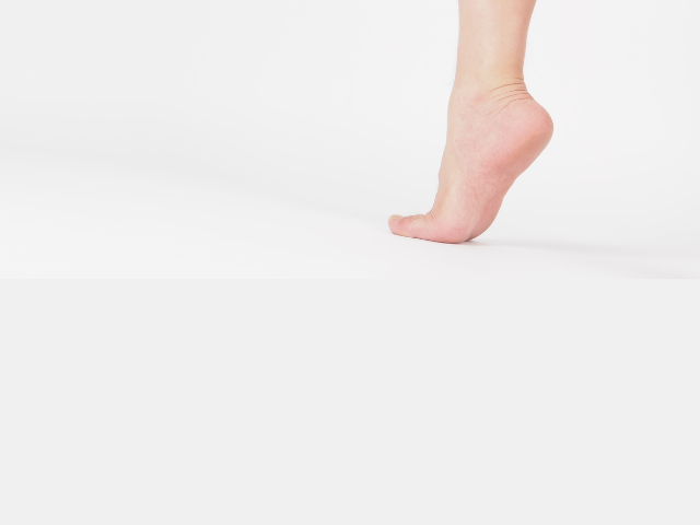
You feel a deep pain in the center of your heel when you walk. This sensation differs from the morning pain typical of plantar fasciitis. It intensifies on hard surfaces. Every step on concrete or tiles makes you wince.
You try to walk on tiptoe to spare your heel. This compensation now creates tension in your calves. The pain persists despite the rest and stretching that usually relieves your foot problems.
You may suffer from heel fat pad syndrome, a condition often confused with other causes of heel pain. However, you must know that this is an uncommon problem even though some healthcare professionals describe a variety of heel diseases with this term.
The heel fat pad, also known as the plantar fat pad, is a specialized structure located under the heel bone, the calcaneus. This layer of adipose tissue organized into fibrous compartments acts as a natural shock absorber. It absorbs impact forces that can reach two to three times your body weight when walking.
Fat pad syndrome occurs when this protective structure deteriorates or shifts. Adipose tissue thins, loses elasticity or migrates laterally. This degradation exposes the heel bone to repeated shocks without adequate protection. The result: a deep and diffuse pain in the heel.
This pathology particularly affects people over 40 years of age, as the pad thins with age. Athletes in high-impact sports (running, basketball, tennis) are also at increased risk. Prolonged use of high-heeled shoes or shoes that minimize cushioning can accelerate pad degradation.
The pain is located in the center of the heel, directly under the heel bone. Unlike plantar fasciitis, which causes sharp pain on the inner edge of the heel, fat pad syndrome creates a sensation of deep bruising or bone bruising.
Discomfort worsens on hard surfaces. Walking barefoot on the floor becomes particularly painful. Prolonged standing intensifies the pain. The direct impact of the heel on the ground — going down a flight of stairs, jumping — triggers sharp pain.
The pain usually decreases with rest and gradually worsens over the course of the day as you accumulate the impacts. This is in contrast to plantar fasciitis, which is typically more painful in the first morning steps.
On palpation, your heel has a marked sensitivity in the center. The fabric may feel thinner or less resilient than a healthy heel.
At Médecine podiatrique du Plateau, Dr. Sandra Gendron, Dr. Stephen Davis and Dr. Émile Carrier, podiatrists, differentiate fat pad syndrome from other causes of heel pain that are much more common.
Clinical examination reveals a specific sensitivity in the center of the heel during direct palpation. We assess the thickness and resilience of the pad by compressing the fabric between our fingers.
X-rays exclude other pathologies such as stress fractures of the calcaneus or heel spurs. Although these bone spurs often accompany plantar fasciitis, their presence does not usually cause the pain of fat pad syndrome.
Musculoskeletal ultrasound visualizes the fat pad, but also the structures in the surrounding area like the plantar fascia attachment on the heel bone. This technology allows your podiatrist to measure its thickness, evaluate its homogeneity and detect associated inflammation or edema. Ultrasound also differentiates this syndrome from plantar fasciitis by examining the adjacent plantar fascia.
Our therapeutic approach aims to protect the heel and optimize the function of the residual pad.
Silicone or gel heel pads redistribute pressure. These insoles cushion impacts and relieve the weakened heel. Different densities and thicknesses adapt to the severity of your problem.
Custom foot orthotics with heel reinforcement provide superior support. We design these devices to maximize damping while maintaining biomechanical stability.
Choosing the right footwear is crucial. Shoes with slightly raised heel (reducing direct impact). Thick, cushioned midsoles. Avoidance of flat or minimalist shoes.
Activity modifications protect your heel during healing, such as temporarily reducing high-impact activities, choosing soft surfaces for activities (grass, treadmill, synthetic track), and gradually progressing as you return to activities.
Cortisone injections can reduce the associated inflammation, but they require special caution. Repeated injection of cortisone into the fat pad may aggravate its atrophy. We reserve this option for refractory cases and limit the number of injections.
Platelet-rich plasma or hyaluronic acid injections are emerging options. These therapies aim to regenerate or increase the volume of the fat pad. While promising, these approaches require more research to confirm their effectiveness.
Dr. Émile Carrier, podiatrist and co-owner of our clinic, explains: “Fat pad syndrome generally responds well to conservative procedures focused on mechanical protection. Patience is essential, as recovery progresses gradually over several months. »
Once the problem has been solved, certain precautions prevent recurrences. Maintain the wearing of well-cushioned shoes. Continuous use of heel pads during demanding activities. Avoidance of walking barefoot on hard surfaces.
Maintaining a healthy weight reduces repeated loads on the fat pad. Each kilogram lost decreases the impact forces on your heels.
If you experience persistent pain in the center of the heel that worsens on hard surfaces, consult for an evaluation. Early diagnosis allows for quick intervention that accelerates your relief.
If you have any questions about heel pain or to make an appointment, contact us at 819 800-1212 or write to us through our contact form.
Podiatric consultations are NOT covered by RAMQ. Private insurance usually covers our services. We will provide you with a receipt that you can submit to your insurer.
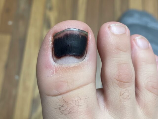
Have you dropped a heavy object on your toe or your nail has been directly impacted during a sports activity and you end up with a black toenail? The pain is immediate and intense. In some cases, the black toenail on the foot occurs after a painless but repetitive microtrauma, for example during running and physical activity with multiple changes of direction such as tennis or pickelball. The toe can hit the toe of the shoe if it is too small or too big.
The following hours reveal the extent of the trauma. A subungual hematoma forms, creating a blue-black spot under the nail. Accumulated blood pressure intensifies the pain. The nail can partially detach from its bed. In some cases, it fractures or tears.
The black toenail is usually the result of a direct impact or repeated pressure on the nail system. This injury can affect the nail itself, the underlying nail bed, or the matrix that produces the nail. The characteristic black or blue-black coloration comes from the blood that accumulates under the nail following the trauma.
The severity varies from a simple subungual hematoma to a complete avulsion of the nail with damage to the surrounding structures.
Nail injuries frequently occur during domestic accidents (heavy object falling on the foot, getting stuck in a door), sports activities (contact sports, running with unsuitable shoes) or repeated traumas (shoes that are too tight, professional activities).
Pain is the dominant symptom, especially when a subungual hematoma creates pressure. You will notice a change in colour under the nail, ranging from red to blue-black depending on the age of the hematoma. The nail can partially or completely detach from its bed. In severe cases, fractures of the distal phalanx (tip of the toe) may accompany the nail injury.
Swelling and tenderness to touch usually persist for several days. If the impact has damaged the nail matrix, the nail that will grow back could have permanent deformations.
At Médecine podiatrique du Plateau, podiatrists Dr. Sandra Gendron, Dr. Stephen Davis and Dr. Émile Carrier carefully assess your nail trauma. The clinical examination determines the extent of the damage. X-rays may be needed to rule out an associated bone fracture.
For painful subungual hematomas, we often perform trepanation. This simple procedure creates a small hole in the nail to drain the accumulated blood. The relief is usually immediate and dramatic.
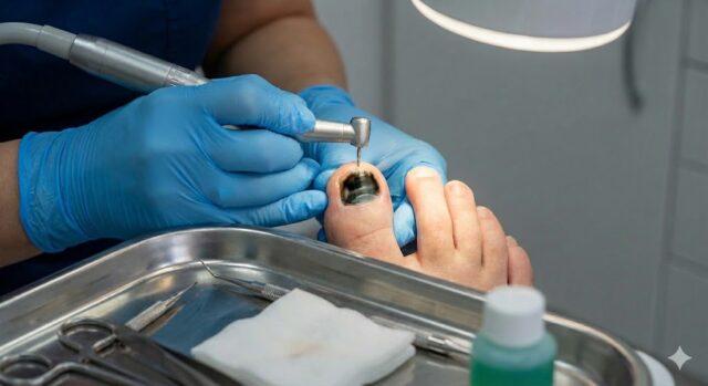
If the nail is partially detached but still attached, we assess whether it can be temporarily preserved as a natural protection of the sensitive nail bed. Completely detached nails are gently removed. If part of the nail is still attached, it may be recommended to remove it to avoid further injury, such as could occur when putting on socks or doing physical activity. In this case, we will recommend a local anesthesia of the toe before proceeding. We then clean and protect the exposed nail bed with an appropriate dressing.
Nail bed lesions sometimes require repair to optimize regrowth. The wounds are cleaned meticulously. Sutures may be required for significant tears.
We also guide you on home care, including protecting the toe during healing, monitoring for signs of infection, pain management and anticipating nail regrowth which typically takes 6 to 12 months for a toe.
Consult a doctor promptly after nail trauma if the pain is severe (especially with a large subungual hematoma), if the nail is partially or completely detached, if you notice a deformity of the toe suggesting a fracture, or if signs of infection appear (spreading redness, warmth, purulent drainage).
Early professional evaluation optimizes healing and minimizes the risk of complications such as infection or permanent nail deformities.
For a quick treatment of your nail trauma, contact us at 819 800-1212 or write to us to schedule your visit.
Podiatric consultations are NOT covered by the RAMQ. Private insurance usually covers our services.

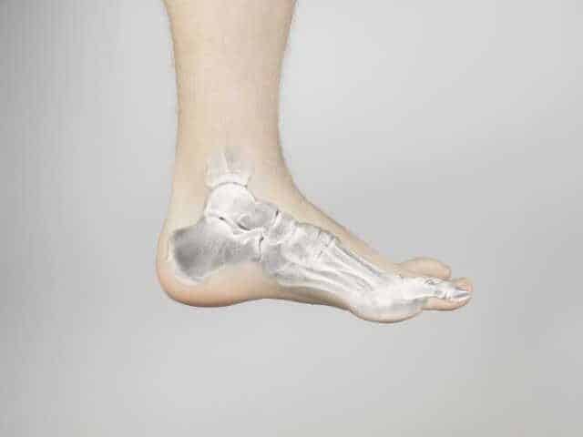
Flat feet are a relatively common condition that affects a large number of people around the world. It is characterized by the absence or collapse of the plantar arch, resulting in a plantar surface that is entirely or almost entirely in contact with the ground. While some individuals may live without major symptoms, others may experience pain, posture disturbances, or difficulty walking, depending on the severity of the problem. This introduction aims to shed light on the potential causes, manifestations, and treatment options to better understand this condition.
Flat foot is a presentation in which the arch of the foot, i.e. the curve formed by the bones and tissues that support the weight of the body, is sagging or absent. In other words, this part of the arch of the foot touches the ground entirely or almost entirely when standing. This characteristic can be present from birth (congenital flat foot) or develop with age due to poor posture or injury to tendons and ligaments as is the case in adult acquired flat foot, also called posterior tibial tendon dysfunction.
According to a study by the American Academy of Orthopaedic Surgeons, about 20-30% of the world’s population has flat feet. This problem can affect one or both arches and may be more common in older adults and those with obesity issues. Although some people live without significant symptoms, flat feet can lead to pain in the feet, ankles, legs, and even the back, as well as postural instability that can cause injury.
Flat feet can be inherited genetically, especially in the case of congenital flat feet. In this case, the bone structure and tissues that support the arch may be defective from birth.
An injury to the foot, ankle, or Achilles tendon can also lead to a collapse of the arch of the foot. In addition, certain diseases such as inflammatory arthritis, neuropathy, or loss of sensation, which can be a complication of diabetes, or neurological problems can cause muscle weakness and consequently lead to flat feet.
Poor posture, obesity, lack of exercise, and wearing inadequate footwear can also contribute to the development of flat feet. Spending a lot of time standing or performing activities that put excessive strain on the feet can also increase the risk of developing this problem.
Symptoms associated with flat feet can vary in intensity and include:
To diagnose flat foot, our podiatrists will perform a physical examination and ask questions about the symptoms experienced. Then, they will do a biomechanical examination, an analysis of gait and standing posture as well as a study of plantar pressures. X-rays may also be used to assess the bone structure of the foot and determine if there is a collapsed arch. Your podiatrist can also do certain clinical tests, for example to assess the strength of certain muscles.
People with flat feet may have difficulty walking or maintaining balance due to the lack of support in their feet. This can lead to abnormal posture and cause pain in other parts of the body, such as the knees, hips, and back.
The lack of stability and misalignment caused by flat feet can increase the risk of ankle, knee, and hip injuries. Athletes and people who regularly engage in physical activity may be particularly vulnerable.
Flat feet can also cause excessive wear and tear on the foot and ankle joints, which can lead to arthritis problems and other long-term joint complications. The big toe joint, which is particularly stressed as a pivot when walking, is at risk of partially blocking in the presence of a flat foot. This often leads to premature wear and tear of the joint, which is called osteoarthritis.
The pain, discomfort, and physical limitations caused by flat feet can significantly affect a person’s quality of life. Daily activities such as walking, running, and even standing for long periods of time can become difficult and uncomfortable.
There is no one-size-fits-all treatment for flat foot, as it will depend on the underlying cause and severity of the symptoms. In some cases, flat feet may not require treatment if the symptoms are mild and do not cause discomfort or interfere with daily activities.
However, in cases where flat foot is more severe and affects a person’s quality of life, there are several treatment options available. Non-surgical approaches such as custom-made foot orthotics, strengthening and stretching exercises, and wearing appropriate footwear can help improve foot stability and reduce symptoms.
In addition to the main treatments, ancillary care such as massage and stretching can also be beneficial in relieving the pain and discomfort caused by flat foot.
In rare cases, surgery may be recommended if symptoms do not improve with other treatments or if the underlying cause of flat foot requires correction. There are different types of surgical procedures available, each with its own advantages and disadvantages. It is important to discuss in detail with your healthcare team. It should be noted that surgery is generally only offered as a last resort if other treatments are not effective. It is not widely used in Quebec and Canada to treat pain associated with flat feet.
Adopting the best practices can make all the difference for people with flat feet. Targeted exercises to strengthen the arch and ankle muscles help support the foot and improve its alignment. During physical activities or when you are on your feet for a long time, be alert to signs of fatigue or discomfort and give yourself breaks to avoid aggravating symptoms. Finally, choosing the right footwear with good arch support and adequate cushioning is key to maximizing your comfort and preventing pain.
In summary, flat feet are a common problem that can be caused by a variety of factors such as genetics, injuries, or certain health conditions.
If you have any concerns about your feet, we advise you to make an appointment for an evaluation with one of our podiatrists. Call us at 819-800-1212 or fill out the contact form to request an appointment.
The foot is a complex and essential structure that supports our body during each step. However, certain types of feet such as cavusfoot, i.e. a foot with a very high arch, can lead to pain, discomfort or posture problems. The purpose of this guide is to explain the causes of cavus foot, its consequences on your health and the diagnostic and therapeutic solutions offered by Médecine podiatrique du Plateau.
The cavus foot is characterized by an overly accentuated plantar arch, unlike the flat foot where the arch is sagging. The causes of cavus foot vary and can include:
It is essential to determine the exact cause in order to offer an appropriate treatment.
If cavus foot is not properly taken care of, it can lead to many problems, such as:
These consequences should not be taken lightly, as they can have a significant impact on quality of life.
At Médecine podiatrique du Plateau, we have advanced tools and technologies to accurately diagnose cavus foot. Here is an overview of the examinations we conduct:
This examination evaluates the functioning of your feet in movement and in a static position. We analyze the distribution of your weight and the way your feet handle the pressures.
Using specialized cameras and a pressure mat, we record and analyze your way of walking. This allows us to detect problem areas and adapt the treatment accordingly.
We offer on-site X-rays, which provide an accurate view of the bone structure of the foot. This is particularly useful for identifying possible deformities or complications related to cavus foot.
These examinations make it possible to establish an accurate diagnosis and develop a personalized treatment plan.
Once cavus foot is diagnosed, we offer different options to relieve your pain and improve your quality of life.
Foot orthotics are insoles that are tailored to your specific needs. They help to redistribute pressure on the foot and improve posture and gait.
Targeted exercises may be recommended to strengthen the foot muscles, improve flexibility, and correct imbalances.
In some cases, cortisone injections or other drug treatments may be offered to reduce inflammation or acute pain.
If conservative treatments are not enough, surgery is considered to correct the deformed or damaged structures of the foot.
Cavus foot can be effectively managed to minimize its impact on your daily life. At Médecine podiatrique du Plateau, we are committed to offering you personalized care based on accurate diagnoses and adapted treatments.
Don’t wait any longer to take care of your feet. Contact us today for a consultation and begin your journey to better podiatric health.

When it comes to foot or ankle fractures, prompt evaluation and proper treatment are essential to ensure complete healing and avoid long-term complications. In this article, we’ll discuss everything you need to know about foot and ankle fractures: how to identify them, why prompt treatment is crucial, and how Médecine podiatrique du Plateau in Gatineau can support you with innovative solutions like X-rays and on-site ultrasound.
A foot or ankle fracture is a partial or complete rupture of one or more bones in these structures. These fractures can occur as a result of direct trauma, such as a fall, or repetitive strain injuries, often caused by sports activities.
The severity can vary depending on the type and location of the fracture, requiring specific management.
Recognizing the early signs of a fracture is crucial to getting timely treatment. Here are some symptoms that should alert you:
If you notice one or more of these symptoms, don’t wait for hours in the emergency room. Make an appointment directly with our clinic at 819 800-1212 for a quick and accurate diagnosis.
Médecine podiatrique du Plateau stands out for its personalized approach and its diagnostic tools available on site. Unlike long waits in hospitals or medical clinics, we offer quick access to essential services to accurately diagnose fractures.
Our services include:
With these state-of-the-art tools, we take care of your condition quickly, without the frustrating delays often encountered in emergency departments.
A delayed or incorrect diagnosis can lead to complications: poor healing, permanent weakness, or even early osteoarthritis. With advanced technologies such as X-rays and ultrasounds, the podiatrists at Médecine podiatrique du Plateau in Gatineau can assess the situation on site, thus avoiding hours of waiting in the emergency room.
Benefits of in-clinic diagnostics:
Obviously, any fracture that is open or that appears to pose a serious health risk should be treated urgently in hospital. It is recommended that you go to the nearest emergency room in such cases.
Immediate and appropriate treatment is necessary to ensure optimal healing and avoid complications such as arthritis or chronic pain. Here’s how we manage fractures:
At Médecine podiatrique du Plateau, care is personalized to maximize your chances of a full return to your activities.
We know that foot or ankle injuries can’t wait. Here’s why our clinic is a great choice if you need help with a possible fracture:
Managing your medical expenses is made easier thanks to the coverage of several services by private group insurance plans, including the Canada Public Service Health Care Plan. We will provide you with a receipt that you can submit to your insurer for reimbursement under the terms of your plan.
Foot and ankle fractures, although bothersome, can be effectively treated with prompt diagnosis and proper management. If you experience any pain or symptoms, don’t wait.
Make an appointment today with Médecine podiatrique du Plateau in Gatineau and benefit from a complete and accessible treatment. Book easily online or by phone at 819 800-1212. Since we want to reduce delays as much as possible, it is best to call the clinic to get the appointment as quickly as possible. Together, let’s get back on your feet.
Médecine podiatrique du Plateau in Gatineau is here to help you regain your mobility and comfort with complete peace of mind. The health of your feet is our priority!
Listen to this video clip from Dr. Émile Carrier, podiatrist, about the 5 common mistakes in the management of nail fungus (onychomyosis) – in French (English subtitles available). It gives you precise and easily applicable advice to maximize your chances of eradicating the problem effectively.

Nail fungus, also known as onychomycosis, is a common fungal infection that affects the nails of the hands or, more commonly, the toenails. Although often considered benign, this problem can lead to complications if not treated promptly. In this article, we will explore the main causes, the importance of treatment, the diagnostic options available at Médecine podiatrique du Plateau, and the specialized care we offer. You will know everything to better understand and act against nail fungus.
Onychomycosis is caused mainly by dermatophytic fungi, but can also result from other pathogens such as yeast or mold. Here are some factors that promote its appearance:
In short, we often get nail fungus for no easily apparent reason.
Many people overlook nail fungus, thinking that it is a purely cosmetic problem. However, its consequences can go far beyond that:
Treating nail fungus in its early stages helps avoid these complications while reducing the risk of recurrence.
At Médecine podiatrique du Plateau, we offer advanced diagnostic services to identify onychomycosis and tailor care to your needs. Steps include:
With an accurate diagnosis, we can offer a suitable care plan, increasing your chances of a quick recovery.
Your health is our priority and our podiatrists provide you with state-of-the-art treatments to get rid of nail fungus. Looking for the miracle trick? In fact, there is not just one cure, but a set of treatments that are part of a complete treatment program that we offer to our patients to solve their nail fungus problem, namely:
Depending on the severity and nature of the infection:
We use specialized techniques to clean and debride infected nails. This procedure reduces the thickness of the nail and removes infected parts, making it easier for antifungals to penetrate. We use quality milling equipment with autoclavable handpieces as well as perfectly sterilized instruments at our sterilization plant in our clinic.
Treating onychomycosis is not instantaneous. Our podiatrists will accompany you throughout your therapy with regular follow-up consultations until the infection is completely resolved. This follow-up includes advice to avoid recurrences:
Nail fungus may seem harmless, but quick and effective treatment is essential to avoid unnecessary complications and discomfort. At Médecine podiatrique du Plateau, our knowledge, know-how and personalized care offer you optimal treatment. Contact us today to schedule a consultation and get your nails healthy.
Make an appointment now and take the first step towards a sustainable solution!
Sesamoiditis is a common foot condition that can cause pain and discomfort in the ball of the foot. As a leading podiatry clinic in Gatineau, we understand the impact this condition can have on your daily activities. In this post, we will explore the causes, symptoms, and treatment options for sesamoiditis, providing you with valuable insights and guidance to help you manage and overcome this condition.
Sesamoiditis is an inflammation of the sesamoid bones, which are small bones located in the tendons of the foot, just beneath the big toe joint. These bones act as a pulley system, helping to provide leverage and absorb pressure during activities like walking and running. However, repetitive stress or injury to the sesamoid bones can lead to inflammation and pain, resulting in sesamoiditis.
Sesamoiditis can be caused by a variety of factors, including:
The most common symptom of sesamoiditis is pain in the ball of the foot, typically focused beneath the big toe joint. The pain may worsen with activities that involve bending the big toe, such as walking, running, or jumping. Other signs and symptoms may include swelling, difficulty bearing weight on the affected foot, and a limited range of motion in the big toe.
At our Gatineau podiatry clinic, we offer a range of treatment options for sesamoiditis, tailored to individual needs and severity of the condition. Treatment may include:
If you are experiencing foot pain or suspect you may have sesamoiditis, we recommend scheduling a consultation with our experienced podiatrists. Our team will conduct a comprehensive assessment, provide an accurate diagnosis, and create a personalized treatment plan to help you find relief and get back to your active lifestyle.
Stay tuned for our upcoming blog posts, where we will dive deeper into common foot conditions, prevention tips, and the latest advancements in podiatry care. For any questions or to schedule an appointment, please contact our podiatry clinic in Gatineau. Your foot health is our priority!

Stress fractures are fine cracks that develop in the bone due to the repeated application of low-level forces. These fractures occur when the bone is subjected to repetitive stresses, such as those experienced during running or other high-impact activities. Stress fractures are common in athletes, especially those who participate in sports that involve repetitive runs or jumps.
At first, X-rays may appear normal, as stress fractures are not always visible.
X-ray: They may eventually reveal a thin line or area of increased density in the affected bone as the fracture heals and the bone callus forms.
Bone scan: This imaging test uses a small amount of radioactive material to detect areas of increased bone activity. Bone scans can detect stress fractures at an early stage, even when X-rays are normal.
MRI: Magnetic resonance imaging is a more sensitive imaging test that can provide detailed images of bone and soft tissue. MRI can show bone edema, periosteal reaction, and the fracture line itself, confirming the diagnosis.
Treatment for stress fractures in the foot is usually non-surgical and aims to reduce pain and allow the bone to heal.
Rest: Avoiding activities that cause pain is crucial to allow the fracture to heal. The duration of the resting period varies depending on the severity of the fracture and its location.
Ice: Applying ice to the affected area can help reduce pain and inflammation.
Medications: Over-the-counter pain relievers, such as ibuprofen or naproxen, can help relieve pain and inflammation. However, make sure you can take it safely. If in doubt, talk to a healthcare professional. In some cases, stronger medications, such as prescription anti-inflammatories or other pain relievers, may be prescribed for severe pain.
Immobilization: In some cases, a cast or walking boot may be needed to immobilize the foot and promote healing. The duration of immobilization depends on the location and severity of the fracture.
Physical therapy: Once the fracture has healed sufficiently, physical therapy exercises may be prescribed to help restore the strength, flexibility, and range of motion of the foot. Physical therapy can also help correct biomechanical abnormalities that may have contributed to the stress fracture.
Proper warm-up: Warming up properly before exercise can help prepare muscles and bones for the stress of activity, which can reduce the risk of stress fractures.
Gradually increase the intensity of the workout: Gradually increasing the duration and intensity of the exercise allows the muscles and bones to adapt to the increased loads, which can minimize the risk of stress fractures.
Appropriate footwear: Wearing supportive footwear that is appropriate for the type of activity can help prevent stress fractures. Replace running shoes regularly, as cushioning wears out over time.
Balanced diet: Eating a diet rich in calcium and vitamin D is essential for bone health. Consult a health care practitioner to determine if calcium or vitamin D supplementation is required.
Stress fractures in the foot are a common injury in athletes and active people. Understanding the causes, symptoms, and treatment of stress fractures can help individuals take steps to prevent these injuries and receive appropriate medical care in the event of a fracture. Early diagnosis and treatment are essential for optimal healing and a pain-free return to activities.

Knee pain can often be caused by misalignments in the foot that affect biomechanics. At Médecine podiatrique du Plateau, our team of podiatrists in Gatineau, Hull and Aylmer use foot orthotics to reduce these imbalances and relieve pain. Here are the main causes of knee pain from the foot:
Pronation is the inward movement of the foot, but when excessive, it causes internal rotation of the tibia. This movement can then overload the knee joint.
Custom foot orthotics can help limit overpronation, reducing strain on the knee.
Supination, which is the outward rolling of the foot, creates lateral instability of the foot and increases impacts on the lateral side of the knee.
Foot orthotics for supination can improve stability and limit the impact on the knee.
Deformities such as a first ray in plantar flexion, i.e. a foot where the first metatarsal (the bone just before the big toe) is pointed downwards, alter the biomechanics of the foot, resulting in compensatory movements in the knee.
Thanks to the right foot orthotics , a podiatrist can correct these imbalances, providing better function of the lower limb.
A problem with the subtalar joint reduces the mobility of the foot and affects the alignment of the knee.
Adapted foot orthotics reduce compensatory foot movements to reduce the impact on the knee.
Even a slight difference in length between the legs can create asymmetry in pronation, putting excessive pressure on the knee. In fact, one of the two feet, usually the one with the longer leg, can collapse in order to reduce the functional length of the leg. Although this does not shorten the leg bones, the more pronounced pronation of one foot can reduce the height of the pelvis on this side, so that the pelvis is better aligned. Since the pelvis is the foundation on which the spine rests, a well-aligned pelvis is very practical!
Compensatory foot orthotics help balance the length of the legs and relieve pressure on the knee.
Knee pain caused by the foot requires a solid knowledge of the biomechanics of the foot, a complex organ of movement. At Médecine podiatrique du Plateau, our podiatrists in Gatineau and Aylmer offer a complete assessment to determine the precise cause of your pain and custom foot orthotics adapted to your needs.
Metatarsalgia refers to localized pain in the forefoot, often in the heads of the metatarsals. This problem, also known as plantar plate dysfunction, can lead to considerable discomfort when walking, exercising, or even resting.
Metatarsalgia can be caused by a variety of factors, including:
Common symptoms include:
At Médecine podiatrique du Plateau, we carry out a complete evaluation to diagnose metatarsalgia. This includes a detailed physical examination, mobility tests, and possibly X-rays to rule out other problems, such as fractures or osteoarthritis.
Treatment for metatarsalgia depends on the underlying cause and may include:
If you are experiencing pain in the front of your foot, it is essential to consult a podiatrist for an accurate diagnosis and an appropriate treatment plan. At Médecine podiatrique du Plateau, our team is dedicated to helping you regain comfort and mobility.

Osteoarthritis of the big toe, also known as hallux rigidus, is a form of osteoarthritis that affects the joint at the base of the big toe. This problem is characterized by stiffness and pain, making foot movements difficult and affecting quality of life.
Hallux rigidus often results from the progressive wear and tear of the articular cartilage, which leads to pain when walking, sports activities, or even at rest. Symptoms may include:
At Médecine podiatrique du Plateau, we perform a thorough clinical examination to assess the pain and mobility of the big toe. Additional tests, such as x-rays, may be done to confirm the diagnosis and assess the extent of osteoarthritis.
Treatment for hallux rigidus can vary depending on the severity of the condition. We offer several options, such as:
If you suffer from big toe pain, it is crucial to consult a podiatrist for an early diagnosis and an appropriate treatment plan. At Médecine podiatrique du Plateau, our team is here to help you manage your symptoms and regain your mobility. Our success is to know that you are on your own two feet, capable of doing all the activities you want with as little pain as possible.
Take this problem seriously. An assessment will allow you to see clearly and evaluate treatment options. Call now at 819-800-1212 to schedule your first appointment.
Did you know that your back pain could be related to foot or posture problems? Podiatry plays a key role in the assessment and treatment of these pains, especially when they are caused by asymmetry in the feet, legs or joints. Indeed, walking is a naturally symmetrical process. As soon as this symmetry is compromised, pain can appear not only in the feet and ankles, but also in the knees, hips, and back.
Here are some examples of disorders that can upset this balance and lead to back pain:
At Médecine podiatrique du Plateau in Hull, Aylmer, Gatineau, we offer an in-depth gait analysis to identify these postural disorders and asymmetries. In addition, our X-ray of the feet (X-rays) allows us to better understand the condition of the skeleton of your feet, bones and joints and to detect any problems that may be contributing to your current problems or future problems. Once the podiatric diagnosis has been made, we offer tailor-made solutions, such as foot orthotics, injections, surgeries, adapted shoes, exercises, physiotherapy or chiropractic treatments to restore balance and relieve your pain.
If you suffer from back pain but don’t know its origin, consulting a podiatrist in Gatineau can offer you a fresh perspective and concrete solutions. In collaboration with other professionals, including chiropractors whose scope of practice includes spinal alignment assessment, your podiatrist can help you relieve your pain.
Take the first step towards relief. Call 819-800-1212 to schedule your first assessment.
You have a bump on your toe. Do this bump make your feet sore when you wear shoes? It could be a hallux valgus, commonly known as a foot bunion.
This condition may be genetic in origin. It can also develop due to our activities, our occupation or the shoes we wear. Thus, you can avoid further aggravation if you undertake changes in your habits and start preventative treatments.
A variety of non-surgical treatments are available at Médecine podiatrique du Plateau. For example, our podiatrists can help you find the shoes best suited to the condition of your feet and the prevention of this problem. Custom-made foot orthotics, which are molded insoles prescribed to alleviate mechanical problems in your feet, are devices often used for bunions of the foot. In addition, your podiatrist may recommend a cortisone infiltration, a digital orthosis or a toe brace.
When non-surgical treatments have been attempted, you have two options for surgery:

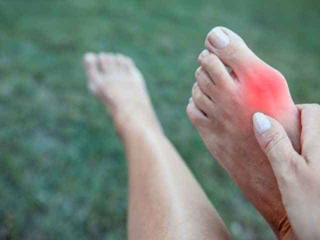
Does your child go to the pool and come back with bumps under their feet? Don’t delay. He may have caught contagious plantar warts. If you are an adult, you should know that this problem can also affect you.
A plantar wart is a lesion on the skin caused by a virus, the papillomavirus or human papillomavirus. Small blackheads and a crust of hard skin or horns can sometimes be observed at the lesion. It is important to recognize and eliminate them as quickly as possible before they grow and spread. This virus is found on the ground and is usually caught by walking barefoot. This is why they are more often found in children. It is therefore advisable to wear sandals, shoes or stockings in public places to avoid contamination. A good cleaning of the shower and bath at home is also recommended.
Grandma’s remedies such as essential oils and duct tape to remove the wart root are not effective. Several products are available in pharmacies. There are salicylic acid dressings (Compound W) and cold treatments (cryotherapy) that try to destroy the wart with liquid nitrogen. However, these types of treatments are often too weak to eliminate the virus completely. Sometimes, their ineffectiveness even causes an increase in the size of warts and the development of new warts.
It is important to make the correct diagnosis and not to confuse your wart with other skin problems such as fungus, blisters, corns or calluses. This could explain the failure of basic treatments and the pain.
The podiatrist will be able to offer you more effective treatments to treat and quickly remove your plantar wart that seems impossible to treat and ensure the correct diagnosis;
The podiatrist will also be able to tell you when the wart virus has died. To do this, he uses a specialized lamp called a dermatoscope. This allows him to see the signs of plantar warts or to notice their absence as if in a magnifying glass.
The clinical examination of your podiatrist during a treatment of nails and skin (foot care) allows to detect and treat plantar warts.
We bring our energy, experience and knowledge to guide our clients towards the solution to their challenges and a more enjoyable life.